 My original post on technologies of human replication, a year ago, was mostly about numbers. I estimated that a ‘fair copy’ of a human being, functionally indistinguishable from the original, could be constructed from approximately 1016 bits of information. That’s a petabyte – a lot of data by today’s standards, but not mind-boggling. One petabyte = 1024 terabytes (Tb). The going price for a 1 Tb hard drive today is $70 retail; 1024 of them would cost $71,680 (less with volume discounts). By Moore’s Law, that cost should drop to 0.7 cents by 2056. In that time-frame, other technologies required for human replication will have matured. As I argued, the business case for developing such technologies is compelling. Three business drivers – applications where the economic benefits justify the required R&D – are transportation, health, and life insurance.
Some implications of the transportation and life insurance applications have already been discussed in the Phantom Self blog. Information-based teleportation –human replication as a means of transportation – is a game-changer for the transportation industry, but will not, by itself, turn society upside down. Travelling to Omaha as a stream of data will quite naturally become the usual way of doing business. The use of this technology for life insurance – restoring people from backups, after they have died – is more radical, because it changes our relationship to death. Death will be transformed from the ultimate loss to a temporary setback, like the setback called ‘death’ in a video game.
I haven’t said much about health applications. But in the medium-term future, health care looks like the biggest driver of all. Health care costs in developed nations are escalating towards fiscal crisis. According to the 2007 Commonwealth Study, the United States spends 16% of its GDP on health care – triple the percentage it spent in 1960. The trend shows no signs of abating. Average age in the developed world continues to rise, and annual health care costs increase with the age of the patient.
We need to do things better. An interesting development in health care technology, directly related to human replication, is organ printing.
Bioprinting – Human Replication, One Organ at a Time
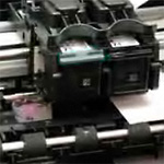
In recent years, the engineered construction of human organs has moved from speculative fiction to clinical reality. Human bladders are now built in labs and surgically implanted in patients. In animal studies, skin patches are constructed layer by layer in vivo, to cover and heal burn wounds. A research project is underway to replicate the human kidney, one of the most complex organs in the body. All these organs are made of cells grown in lab cultures originating from stem cells extracted from the patients’ bodies. One of the technologies enabling this remarkable progress is the home-and-office inkjet printer.
At Wake Forest University’s Institute for Regenerative Medicine, Anthony Atala leads a team of seven hundred researchers building a variety of body parts including hearts, livers, kidneys, muscles, arteries, skin, ears, fingers. Bioprinting using ink-jet technology is just one of the team’s techniques.
Atala made history when his biologically engineered bladders were the first laboratory-grown organs ever used in human trials. Beginning in 1999, several young children with spina bifida received bladders grown from their own cells. The patients are still doing well. The bioengineered bladders perform better than bladders repaired with surgically transplanted bowel tissue, using an older technique.
The bladders were not produced by inkjet printers, but by an older technique requiring a ‘scaffold’. The process began by extracting a few cells from the patient’s own bladder tissue. The cells were cultured in glass dishes containing a growth medium, where, over a month, they reproduced to the required numbers (about 1.5 billion). They were then applied to a bladder-shaped scaffold made of collagen, a natural material found in the structural framework of most organs. A collagen scaffold can be made by using a “mild detergent” to chemically remove all cells from a donated organ. Because the collagen is non-cellular, and is biologically indistinguishable from one person to another, it can be transplanted without fear of rejection. After maturing for several weeks, the bladders were surgically transplanted to the patients. Because each patient received a bladder made exclusively from his own cells, his or her immune system did not reject the transplanted organ.
Organs built on collagen scaffolding were the state of the art in bioengineering at the turn of the millennium. For human use, the bladder application has been one of the most successful so far. Experiments on animals have taken more exotic turns.
As far back as 1995, Massachusetts researchers Vacanti and Griffith-Cima reshaped bovine cartilage in the form of a human ear and implanted it under the skin of a hairless mouse. The purpose was to demonstrate fabrication of cartilage structures, with a view to human reconstructive surgery. Because the mouse belonged to a immunocomprimised strain commonly used in experimentation, it did not reject the transplanted bovine cells. Pictures of the “ear-mouse” still haunt the internet, provoking outraged calls for curbs on the excesses of science.
The field of tissue regeneration should not be judged by an occasional marketing blunder. Much legitimate, valuable progress is being made. Yale researchers led by Laura Niklason created a scaffold by decellularizing a pair of rat lungs, then seeded it with lung cells from a second rat. When the organ matured, it was surgically implanted into the second rat. “The lungs breathed in the new rat and exchanged air for up to two hours” (after which they were removed for analysis). The technique of growing organs on scaffolds extracted from cadavers holds great promise for increasing the supply of much-needed human transplant organs.
Gabor Forgacs, at the University of Missouri, used bioprinting to create functioning chicken heart tissue in 2006. A bioprinter was used to spray layers of tiny clumps of cells into a petri dish, in the desired shape. Layers of cells were alternated with layers of a supporting gel, called “biopaper”. Adjacent cells fused together. Cells printed in rings, in many layers, fused to form tubes which could function as a blood vessels. Not only did the cells fuse, but most importantly, cells printed together in this way began to work together as they would in a natural organ. Nineteen hours after being printed, the chicken heart tissue started “to beat in a synchronous manner.” A 2009 TED talk by Atala shows “a typical desktop printer”, its print-head packed with cells instead of ink, printing a two-chamber heart in forty minutes. “Four to six hours later you see the muscles contract.”
Such rapid progress in tissue engineering is helped by the fact that bioprinting techniques harness natural processes of cellular self-assembly. As explained in the 2010 paper Tissue engineering by self-assembly and bio-printing of living cells by Jakab et al, molecular properties of cell walls – properties like surface tension – induce automatic sorting and fusion of living cells. Kept alive in a bioreactor (an environment maintaining normal body temperature and supplying oxygen), a liquid mixture of various cell types will self-assemble into solid tissue. Self-assembly only works at a fine level of detail; but that, after all, is where most of the work must take place. Larger structures – layers of different cell types, the vascular network – can be shaped directly by the printing process. The promise of bioprinting rests largely on assistance from natural processes: first, in reproducing cells to yield the numbers required to build organs, and second, in assembling those cells into tissue.
Scaffold-free bioprinting techniques are now being used to build the vascular tree, including small blood vessels, required for the internal blood transport that is essential to keeping all but the thinnest organs alive. In a 2009 paper, Scaffold-free vascular tissue engineering using bioprinting, authors Norotte, Marga, Niklason, and Forgacs describe the benefits of the process:
A unique aspect of the method is the ability to engineer vessels of distinct shapes and hierarchical trees that combine tubes of distinct diameters. The technique is quick and easily scalable.
Ink-Jet to Bio-Jet
Dedicated bioprinters have recently been developed to improve on off-the-shelf inkjet technology. Some feature two print heads: one to print droplets of cells, called “bio-ink”, the other to simultaneously lay down a supporting structure of hydrogel, or “bio-paper”. A two-head printer can, in theory, build a large, complex organ without relying on a pre-shaped collagen scaffold. Organovo in the U.S., and Neatco in Canada offer commercial bioprinters; the price of an Organovo unit is in the $200,000 range. For the time being, its customers are labs doing research in tissue engineering; but Organovo’s vision extends to providing printers for clinical organ production – perhaps to print arteries for bypass surgery as soon as 2015, according to CEO Keith Murphy.
Other advantages of dedicated bioprinters include a higher survival rate of printed cells, the ability to print larger clumps of previously aggregated cells, and even to print pre-formed multi-cellular cylinders (i.e. tubes, such as blood vessels) up to 7 cm long. Printing with tubes instead of spheroids results in faster post-printing fusion, as well as more uniform tube diameters.
These techniques offer high precision. According to a recent report, a temporal-mandibular joint (TMJ), which connects the jaw to the skull, was built by a Columbia University team headed by Gordana Vunjak-Novakovic. The process used 3D digital imaging to capture the shape of an existing bone. A scaffold of precisely matching shape was then constructed by an automated process, and infused with bone marrow cells. After five weeks in a bioreactor, the structure became living bone. The Columbia team claims a first in precise bioengineering of a human bone on this scale.
Plenty of challenges, both technical and regulatory, remain before a full suite of complex organs will be available for transplant into human patients. But the research community is not shy about taking them on, and their mood is optimistic. A group led by Vladimir Mironov at the Medical University of South Carolina (MUSC) has embarked on a project to build a human kidney, perhaps the most challenging structure in the human body to replicate (below the neck). The kidneys are responsible for regulating the blood’s pH and maintaining the correct balance of many vital chemicals. Kidney tubules, where blood is cleansed, contain 14 different cell types. The kidney has a complex internal structure, and is tightly coupled to other bodily functions; it is essentially integrated to the blood circulatory system, and its function is regulated by the sympathetic nervous system.
Mironov is undaunted by this complexity. He is quoted in a 2006 report:
The first big step in building an organ requires a plan — a detailed CAD model of it. “I’ve asked anatomists for a digital kidney model with the x, y, and z position of every cell. But they don’t have one, because, they say, no one has ever asked for one. Still, there are sophisticated, noninvasive ways to capture such medical information. MRIs, for example, have resolutions of about 2 mm, enough to see the large branching of blood vessels. But such maps are still just gross anatomy.”
Mironov realized that it is unnecessary to try to replicate a patient’s original kidney on a cell by cell basis. That level of detail doesn’t matter. Instead, organ construction can take advantage of the structural repetition which is characteristic of all biological systems. Mironov’s MUSC website says:
[C]onsidering that organs have a polymeric structure and consist of repeating structural functional units, one can reconstruct only one or two typical organ units and then assemble the whole organ in silico by adding a reconstructed unit based on the gross anatomical structure or by filling the available space. A third approach is based on a mathematical computational anatomical model. For example, by knowing the mathematics of vascular branching it is possible to reconstructed in synthetic materials a very realistic model of the vascular tree found inside the organ. In fact, several commercially available pieces of software permit the creation of a realistic anatomical model from bioimages….
Mironov is confident that bioprinting of functional kidneys suitable for transplant into human patients is technically achievable. When asked for a time-line, he hazarded the date 2030 – but, he stressed, it is not som much a function of time as of funding.
Existing investments devoted to bioengineering technology are enough to fuel a lot of interesting work. In 2009, the National Science Foundation awarded Mironov’s team a $20M grant in support of the kidney project. This is a small fraction of Mironov’s $1B estimate of the total costs of bringing bioprinting kidneys to market. However, the cost is justified by business logic. Harvard’s Professor Joseph Bonventre estimated the market for artificial kidneys at $15B. The bioengineering field is currently supported by large public expenditures such as the $3B committed by California’s Proposition 71 to the Institute for Regenerative Medicine.
To sum up, there is a promising path leading to bioprinting of fully functional organs on a timeline of one to three decades. The primary application will be replacement of damaged or diseased organs by healthy new bioprinted ones, using the patients’ own cells, replicated in a growth medium, as building blocks. This technique exploits the extremely high levels of redundancy in living organisms, and the accuracy of the natural biological processes of cell reproduction. Preservation of detail at the grain size of individual molecules is, for the most part, unimportant. What matters is to produce a functioning organ, of the right size, shape and working capacity, which will not be rejected by the recipient’s immune system. In most cases, replacement organs will have higher functional capacity than the organs being replaced; after all, low functional capacity (usually caused by damage or disease) is the reason they are being replaced. What’s important is that the individual cells of the replacement organ are healthy and functional; that the organ is properly vasculated by a network of veins and arteries to keep its cells supplied with oxygen and nutrients, and to remove waste products; and that the organ’s vascular and nervous systems be properly connected to the rest of the organism. All this is within reach using the three techniques of (1) in vitro replication of the patient’s cells (2) bioprinting of the organ, and (3) surgical replacement of the damaged organ with the healthy bioprinted one.
The technology described in this post will not directly lead to the kind of information-based human replication required for teleportation and life insurance. It does not address the problem of replicating the informational content of the brain. It requires at least some physical cells from the human subject as starting points for replicating the cells needed to construct the different organs. However, the bioprinting assembly techniques being developed today, once speeded up, could be used as part of the human replication process. The ability to exploit cellular self-assembly – getting an assist from nature – is another enormous advantage, one that, surprisingly, may make it easier to automate the replication of living organisms than of inanimate objects of similar complexity. And reliance on structural repetition within the human body will enable data compression, vastly reducing the amount of information needed to reconstruct everything that matters about a person.
Today’s bioengineering agenda holds another, perhaps even more alluring promise: bodily rejuvenation by full body transplant. By ‘body transplant,’ I mean an operation in which a person’s brain is removed from an aging, ‘used’ body and surgically transferred to a youthful, bioengineered body, factory-fresh and defect-free. Bioprinting technology could, within the lifetime of many of us, realize the dream of the Fountain of Youth.
That development is rife with moral implications, not all of which are savoury. It raises questions such as: What would be the social effects of prolonging human lifespan indefinitely? What are the implications for population growth on a resource-limited planet? Would the ability to hold death at bay impede renewal, creating cultural and economic stagnation? Is it fair to younger generations, if older ones refuse to die? It is a development we should start thinking about before it overtakes us.
My speculations about whole-body transplants are no wilder than the visions of active researchers in bioengineering. On the MUSC website, Mironov wrote about creating completely new artificial persons:
If we are able to bioprint all human organs, then by logical extension we will also be able to print an entire human body. What is probably most surprising is that the idea of human printing or engineering of a living human body is not novel. This idea first time came to the human mind at least a thousand years ago. I am talking about Greek mythology. A most famous Greek myth is the story about Pygmalion and Galatea. Shortly, it is a love story about how a sculptor fell strongly in love with the sculpture of a young woman…he created. A Goddess, taking pity, transformed his sculpture into a living girl whom he later married. Bioprinting of human bodies some day could transform this beautiful Greek mythology story into reality.
Reproducing the Brain
A notable exception to the list of organs currently targetted for bioengineering is the brain. One reason for this is that in the brain, unlike other organs, fine detail does matter. The outlook for brain replication will be the topic of the next post.
Click here to view other posts on the Phantom Self home page.

I think that you underplay the hubris of implanting a mouse with a human ear by calling it a ‘marketing error’. The idea, indeed, the pictures I have seen of the poor mouse send shivers down my spine.
I certainly agree that the moral implications need to be considered. We are rushing headlong towards immortality. I wonder what religious people will think of this? Immortality was to be a reward for being good. Soon it will be something that can be purchased with a credit card.